
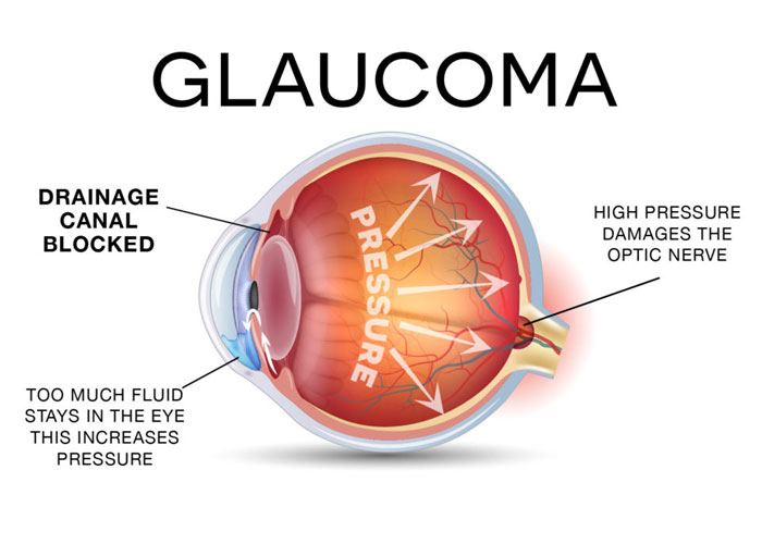
Glaucoma is the term used to depict various related conditions that cause harm to the optic nerve, which transmits data from the eye to the mind. It normally (yet not generally) is connected with high intraocular Pressure. Left untreated, Glaucoma can result in lack of sight.
Glaucoma is an illness of the eye which displays a normal optic neuropathy which brings about dynamic visual field loss. The most essential danger variable is raised intraocular pressure. Risk variables for Glaucoma incorporate age over 40 years, diabetes, nearsightedness, full grown waterfall, trauma, certain retinal maladies and a family history of Glaucoma.
Applanation Tonometry is as of now acknowledged to be the most precise clinical system for measuring the IOP. This strategy in a roundabout way measures the IOP by gaging what amount of energy it takes to smooth the cornea over a settled surface region. This might be similar to pressing the palm of your hand against the surface of an inflatable. On the off chance that the blow up does not have much air in it and it is delicate, then it doesn't take much constrain to blanket the palm of your hand with the surface of the inflatable? In the event that the inflatable has a ton of air in it and it is hard, it will take more constrain to blanket the palm of your hand with the blow up. The expression applanate signifies "to straighten".
The settled surface region is the level tip of the tonometer head. Weight is connected to the cornea through the tip by turning the dial on the tonometer, which discharges weight from a spring inside the tonometer as the dial is moved at the higher numbers. The altered surface range of the tip is secured by the corneal surface when within ring of the mires only touch as saw through the visual of the opening lamp.
Preference of this technique is that it is mounted on the opening light magnifying lens, giving a stable base from which to handle the instrument. Nonetheless, the opening light mount is a burden in that the instrument is not convenient. An alternate weakness is that fluorescein color and a topical soporific must be ingrained into the eye. The fluorescein color helps in review the mires and the soporific is required in light of the fact that the tip touches the cornea.
Gonioscopy is particularly important in instances of pseudo exfoliation, pigmentary glaucoma, tumors or past trauma. In any case, it’s critical to do it on the sum of your glaucoma patients on the grounds that you have to recognize what a solid point looks like so as to diagnose an infected plot. Besides, you would prefer not to be corroded when gonioscopy all of a sudden turns into a key some piece of a case.
Gonioscopy is one of the all the more testing eye examination systems for ophthalmology inhabitants to take in. Lamentably, in clinical practice gonioscopy is not executed as frequently as it ought to be. In an investigation of 193 essential open point glaucoma outlines from a group based example in the United States, just 51.3% of patients had gonioscopy performed at starting presentation (Hertzog LH, Albrecht KG, Labree L, Lee PP. Ophthalmology 1996; 103:1009-13). Gonioscopy is a basic some piece of the eye examination. There is much to be seen by gonioscopy – not basically whether the iridocorneal point is open or closed.
The Visual Field is measured by perimetry. This may be dynamic, where purposes of light are moved inwards until the onlooker sees them, or static, where purposes of light are flashed onto a white screen and the spectator is asked to press a catch in the event that he or she sees it. The most well-known border utilized is the robotized Humphrey Field Analyzer and Heidelberg Edge Perimeter.
An alternate system is to utilize a campimeter, a little gadget intended to measure the visual field.
Examples testing the focal 24 degrees or 30 degrees of the visual field are most normally utilized. Most borders are likewise equipped for testing the full field of vision.
An alternate technique is for the professional to hold up 1,2, or 5 fingers in the four quadrants and focus of a patient's visual field (with the other eye secured). On the off chance that the patient can report the amount of fingers appropriately as contrasted and the visual field of the expert, the ordinary outcome is recorded as "full totally fingers" (regularly shortened FTCF). The blind side can additionally be surveyed through holding a little red protest between the expert and the patient. By analyzing when the red article vanishes for the expert, a patient's strangely huge blind side could be distinguished.
The laser iridotomy is performed in the laser room at the Ambulatory Surgery Center. You don't have to go to the working room. You don't require any blood work or a therapeutic exam before the methodology. You will be wakeful; however your eye will be numbed. The laser system is concise – it takes between 1-2 minutes for the whole strategy. There are no limitations on your exercises after the technique.
Your vision may be blurry for a couple of minutes after the strategy; however your vision ought to return right away.
Laser fringe iridotomy is a medicine for tight plot glaucoma. The specialist utilizes an Nd: yag laser to make a little opening in the fringe iris. This enhances the dissemination of liquid inside the eye and enlarges the foremost chamber point. Liquid which is handled behind the iris has less demanding access to the eye's inner seepage framework. At times this brings down the intraocular weight; however that is not the essential objective of laser fringe iridotomy. The essential objective of the strategy is to decreases the danger of intense edge conclusion glaucoma.
 +91 9509701200
 Mon - Sat: 9:30am - 2:00pm, 5:00pm - 8:00pm, Sunday: By Appointment