
Vitreoretinal eye surgery includes a group of procedures performed deep inside the eye's interior with lasers or conventional surgical instruments.
As the name implies, this delicate surgery takes place where the gel-like vitreous and light-sensitive membrane (retina) are found.
Various vitreoretinal surgical and laser approaches can restore, preserve and enhance vision for many eye conditions such as certain types of age-related macular degeneration, diabetic retinopathy, diabetic vitreous hemorrhage, macular hole, a detached retina, epiretinal membrane and CMV retinitis.
Conditions Requiring A Vitrectomy; How The Procedure Works
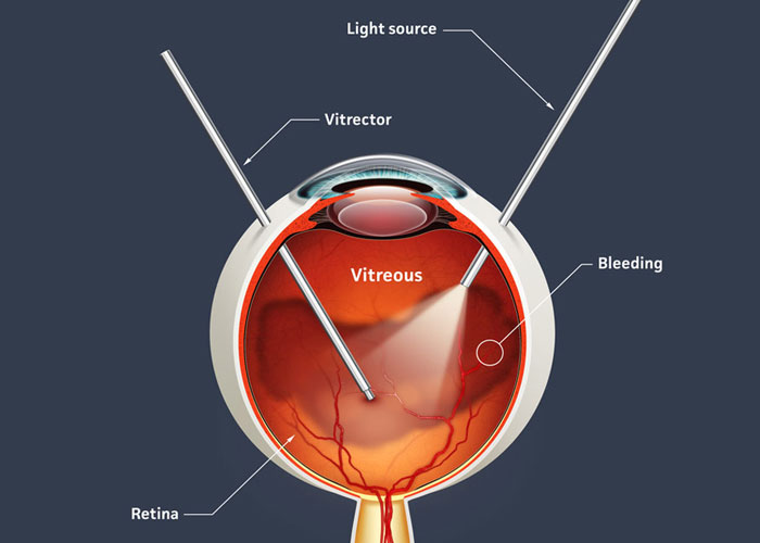
A vitrectomy procedure removes the vitreous humor or gel-like substance in the eye. This approach can address vision problems caused when foreign matter invades this usually pristine area of the eye's interior. One example of foreign matter is blood, from conditions such as diabetic vitreous hemorrhage.
Light rays passing through the eye cause the foreign matter to cast shadows on the retina, resulting in distorted or greatly reduced vision.
Once the surgeon removes the vitreous humor and clears the area, he or she usually injects a saline liquid to replace the vitreous humor that ordinarily fills up the inner chambers of the eye.
However, a vitrectomy is considered inappropriate and extreme for addressing most ordinary spots and floaters that occur with vitreous detachments
affecting almost everyone to some degree as they grow older.
The most common reasons for a vitrectomy include:
Usually vitrectomies require general anesthesia. However, local anesthesia is used in certain situations, especially when general anesthesia would be inappropriate, such as for people with breathing problems.
Your surgeon will make three tiny incisions in the eye to create openings for the various instruments that will be inserted to complete the vitrectomy.
These incisions are placed in the pars plana of the eye, located just behind the iris but in front of the retina. The instruments that pass through these incisions include:
 +91 9509701200
 Mon - Sat: 9:30am - 2:00pm, 5:00pm - 8:00pm, Sunday: By Appointment